Advanced Imaging Concepts in Echocardiography
What will you learn in this module?
Advanced Doppler
Gain a deeper understanding of Colour Doppler, Pulsed Wave, and Continuous Wave Doppler to assess detailed blood flow patterns and cardiac function.
Harmonic Imaging
Learn how harmonic frequencies enhance image clarity, improving the accuracy of cardiac assessments and diagnostic precision.
Speckle tracking & Strain imaging
Discover techniques to measure myocardial deformation, providing insights into heart muscle function and aiding in the early detection of dysfunction.
Complimentary assessments
Understand the role of contrast agents in visualising blood flow and structures and learn about Transesophageal Echocardiography (TOE) for comprehensive cardiac imaging.
Colour Doppler
Colour Doppler uses real-time color-coding to visualize both the direction and speed of blood flow within the heart and vessels. It works by assigning colors to represent the movement of blood relative to the transducer (the ultrasound probe). Typically, red indicates blood flow moving towards the transducer, while blue represents blood flow moving away from it. A good way to remember this is the abbreviation ‘BART’ – blue away red towards.
The intensity or shade of the color also reflects the velocity of blood flow: brighter shades signify higher speeds, while darker shades represent slower speeds. This instant visual feedback allows clinicians to assess blood flow patterns, detect areas of turbulence, and evaluate valve function with ease. For example, abnormal flow patterns or turbulent areas in the color display may suggest conditions like valve leakage or vessel narrowing, helping with early diagnosis and treatment planning.
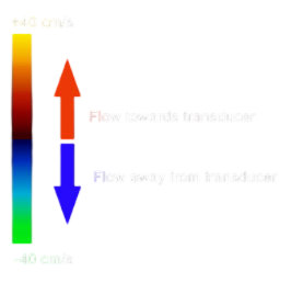
Application in Echo
Colour Doppler provides real-time visualisation of blood flow to assess valve function, detect abnormalities, and identify turbulence. By colour-coding blood flow, it helps clinicians see if valves open and close properly or if blood leaks backward (regurgitation), indicating issues like valve insufficiency or stenosis.
Abnormal flow patterns or turbulent areas (shown as mixed colours or mosaic patterns) can suggest structural problems or obstructions. This quick visual feedback enables accurate, immediate assessments of heart and vessel function.
Continuous wave
Continuous Wave (CW) Doppler is a technique used in echocardiography to measure high-velocity blood flow within the heart and vessels. CW Doppler works by continuously sending and receiving sound waves along a fixed line, allowing it to capture the speed of blood flow without the limitations of pulse duration or depth. This continuous transmission and reception enable CW Doppler to measure high-velocity flows accurately, making it ideal for assessing blood flow through narrowed (stenotic) valves or areas of suspected regurgitation in the heart.
Application in echocardiography: CW Doppler is essential for cases where blood flow velocities exceed the limits that Pulsed Wave (PW) Doppler can measure. For example, when blood flows rapidly through a narrowed valve, CW Doppler can quantify the exact speed, aiding in the diagnosis of valve stenosis.
Comparison with Pulsed Wave (PW) Doppler:
- Benefits of CW Doppler: CW Doppler can measure unlimited velocities, making it particularly useful in high-velocity situations.
- Limitations: Unlike PW Doppler, CW Doppler does not provide precise depth information, as it measures all velocities along the beam path. This lack of spatial resolution means it cannot pinpoint the exact location of high-velocity flow.
Overall, CW Doppler is invaluable for evaluating conditions with high-speed blood flow, while PW Doppler remains preferred for assessing specific locations at known depths within the heart.
Pulsed wave
Pulsed Wave (PW) Doppler is a technique in echocardiography that measures blood flow velocity at a specific location within the heart or blood vessels. It works by emitting sound waves in short, controlled pulses, allowing the ultrasound machine to target a precise depth. The transducer sends out a pulse and then waits for the returning echo, using the timing to determine the distance of the moving blood cells. This selective approach provides detailed information about blood flow at a defined site.
In echocardiography, PW Doppler is commonly used to assess blood flow within specific heart chambers, across valves, or in vessels. For example, it is ideal for measuring peak blood flow velocities through the mitral or tricuspid valves, making it useful in diagnosing conditions like valve stenosis (narrowing) or diastolic dysfunction (impaired filling of the heart).
Harmonic Imaging
Harmonic Imaging is an advanced ultrasound technique that uses harmonic frequencies to create clearer and more detailed images. When an ultrasound wave is sent into the body, it travels at a certain frequency. However, as it moves through tissues, it generates harmonic frequencies—essentially, these are multiples of the original frequency, which provide additional detail in the reflected sound waves.
In harmonic imaging, the ultrasound machine “listens” for these higher, harmonic frequencies rather than the initial, lower frequency. This helps reduce background noise and artifacts (unwanted reflections), resulting in sharper, more accurate images. This clarity is especially useful in echocardiography, where subtle details of the heart’s structure and function are crucial for diagnosis.
Speckle tracking & Strain analysis
Speckle tracking and strain analysis are advanced echocardiography techniques that provide a deeper understanding of heart muscle function. Unlike traditional methods that rely on ejection fraction to assess the heart, these tools allow for precise measurement of myocardial deformation and motion. By tracking natural patterns, or “speckles,” within the heart muscle on ultrasound images, speckle tracking offers detailed insights into how the heart contracts and relaxes in different directions. Strain analysis quantifies this movement, measuring the extent of stretching, shortening, or thickening of the myocardium during the cardiac cycle.
These techniques are particularly valuable in detecting subtle abnormalities that might go unnoticed with conventional imaging. They have revolutionized the evaluation of heart function, offering clinicians an early and more accurate tool for diagnosing and managing cardiac conditions.
Speckle tracking
Speckle tracking involves the identification and tracking of natural acoustic markers, or “speckles,” within the myocardium on ultrasound images. These speckles are unique patterns created by the interaction of ultrasound waves with cardiac tissue, and they remain relatively stable across consecutive frames of an echocardiographic study. Using specialized software, the motion of these speckles is tracked frame by frame throughout the cardiac cycle.
This process allows for precise measurement of heart muscle movement in three directions:
- Longitudinal: Movement of the myocardium along the long axis of the heart (base to apex).
- Radial: Thickening and thinning of the myocardium as it moves inward and outward.
- Circumferential: Shortening of the heart muscle around its circumference.
By analyzing the displacement of speckles in these directions, speckle tracking provides detailed information about regional and global myocardial function, offering an advanced tool for assessing heart health.
Strain analysis
Strain quantifies the deformation of the myocardium by measuring the percentage change in length, thickness, or shape of the heart muscle during the cardiac cycle. It reflects how much a segment of the myocardium shortens, lengthens, or thickens relative to its original size, providing a detailed assessment of myocardial mechanics.
For example:
- Shortening (negative strain) occurs when the myocardium contracts.
- Lengthening (positive strain) occurs during relaxation or stretching.
- Thickening reflects the radial component of strain, indicating how the heart muscle bulges inward during contraction.
The most clinically used strain parameter is Global Longitudinal Strain (GLS), which evaluates the overall shortening of the left ventricle along its long axis (base to apex). GLS is highly sensitive for detecting subtle myocardial dysfunction, often before changes in ejection fraction occur.
Clinically, GLS is a reliable marker for assessing left ventricular function, especially in conditions like:
- Early detection of heart failure.
- Monitoring cardiotoxicity during chemotherapy.
- Evaluating ischaemic heart disease by identifying regional wall motion abnormalities.
GLS has become a cornerstone of advanced cardiac imaging due to its accuracy, reproducibility, and ability to identify myocardial dysfunction early, supporting timely interventions and improved patient outcomes.
Complimentary techniques
Complementary techniques in echocardiography enhance the diagnostic capabilities of standard ultrasound imaging by providing additional detail and specialised insights. These methods, such as transesophageal echocardiography (TEE), contrast echocardiography, and 3D imaging, work alongside traditional transthoracic echocardiography to address specific clinical questions. By improving visualization and diagnostic precision, complementary techniques play a vital role in assessing complex cardiac conditions and ensuring more comprehensive evaluations.
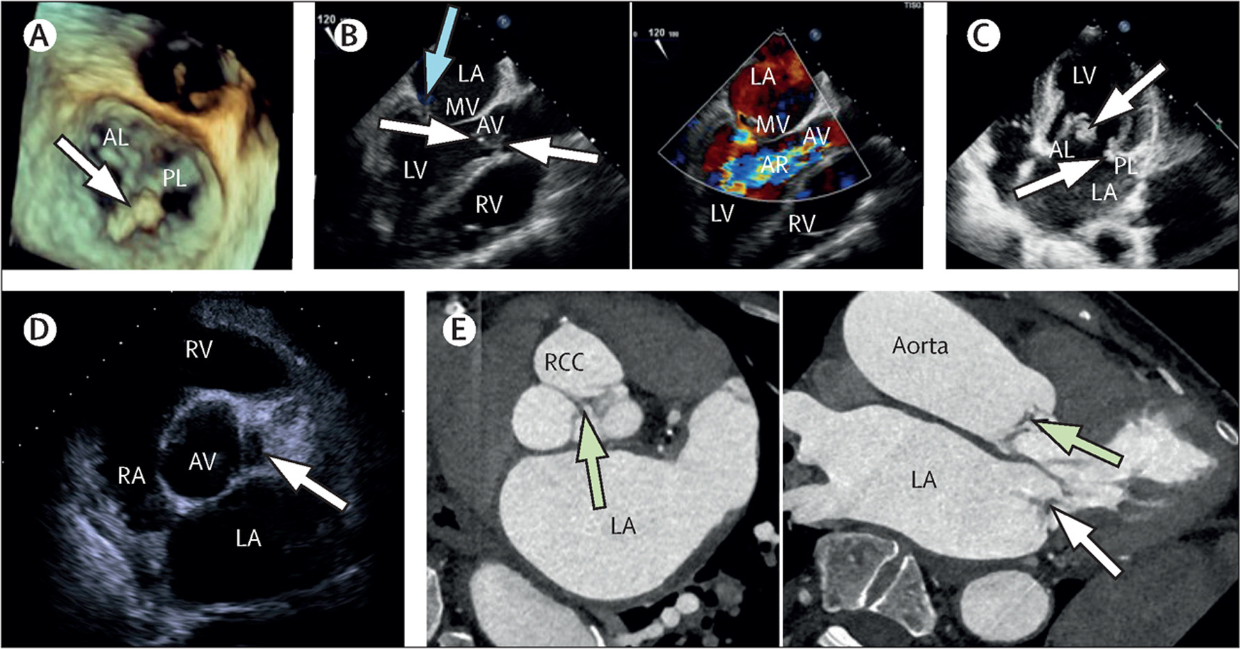
Transesophageal echo
Transesophageal Echocardiography (TOE) provides detailed images of the heart by placing an ultrasound probe in the oesophagus, which lies close to the heart. This proximity allows for clearer imaging of structures, particularly the posterior heart, without interference from the chest wall or lungs.
When is it used? TOE is often used in cases where standard transthoracic echocardiography (TTE) is limited, such as:
- Detecting blood clots, tumours, or infections (e.g., endocarditis).
- Assessing valve abnormalities or prosthetic valves.
- Guiding procedures like mitral valve repair or atrial septal defect closure.
- Evaluating suspected aortic dissection or left atrial thrombus.
Contrast echo
Contrast echocardiography involves injecting microbubble contrast agents into the bloodstream to enhance the visualization of heart structures and blood flow. The microbubbles reflect ultrasound waves, improving the clarity of heart chamber borders and highlighting blood flow patterns.
Stress echo
Stress echocardiography combines ultrasound imaging with physical exercise or pharmacological stress to evaluate the heart’s function under increased workload. It measures how well the heart pumps blood and identifies areas with reduced blood flow or abnormal motion during stress.