Basic principles of Ultrasound Physics
What will you learn in this module?
What is Ultrasound and the nature of Sound Waves
Piezoelectric Effect and Beam Formation
Near Field, Far Field and Image Formation
Doppler Effect and Clinical Applications
What is Ultrasound?
Ultrasound is a medical imaging technique that uses high-frequency sound waves to create detailed images of structures inside the body. These sound waves are emitted from a probe and travel through the body, bouncing back when they hit tissues or organs. The time it takes for these echoes to return to the probe is crucial—by measuring this timing, the machine calculates the distance of the structures, which helps form a real-time image. Safe, non-invasive, and radiation-free, ultrasound is an invaluable tool for diagnosing and monitoring various medical conditions, especially in cardiology and prenatal care.
The nature of Sound Waves
- Frequency
Frequency refers to the number of sound wave cycles per second, measured in Hertz (Hz). In echocardiography, higher frequencies produce better image resolution but have limited penetration depth, ideal for viewing shallow structures like the heart.
- Wavelength
Wavelength is the distance between two consecutive peaks of a sound wave. Shorter wavelengths, which correspond to higher frequencies, result in clearer images but can’t penetrate deeply. Longer wavelengths allow deeper tissue visualization but with lower resolution.
- Amplitude
Amplitude represents the strength or intensity of the sound wave. In echocardiography, higher amplitudes result in stronger echoes, which are important for differentiating between various tissue densities and ensuring clear imaging.
- Speed
Speed is the rate at which sound waves travel through a medium. In echocardiography, sound waves move faster in denser tissues. The speed affects how quickly the image is formed and how accurate the distance measurements are for cardiac structures.
The Piezoelectric Effect
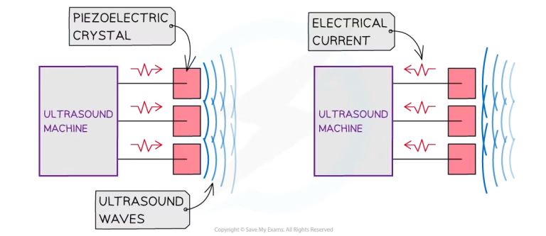
The piezoelectric effect is the key mechanism behind how ultrasound transducers work. Inside the transducer, there are small crystals (usually made of materials like quartz or ceramic) that have special properties. When an electric current is applied to these crystals, they vibrate and produce sound waves—this is the first part of the piezoelectric effect.
In simple terms, the transducer sends these sound waves into the body. As the waves travel through tissues, they bounce back when they hit different structures like organs or blood vessels. These returning sound waves are picked up by the same crystals in the transducer. When the sound waves hit the crystals, they vibrate again, generating a small electric signal. This process is the reverse of what happened earlier.
The ultrasound machine then takes these electrical signals and converts them into images that we can see on the screen. The timing of when the sound waves return helps the machine figure out how far away each structure is, allowing it to form a clear image of what’s happening inside the body.
- Send sound waves out: Crystals vibrate and send sound waves into the body.
- Receive returning waves: Crystals vibrate again when sound waves bounce back, creating electrical signals.
- Convert to images: The machine turns these signals into pictures of what’s inside the body.
Ultrasound transducers
- What is a Transducer?
A transducer is a crucial component of the ultrasound machine, responsible for both sending and receiving sound waves. It converts electrical energy into sound waves using the piezoelectric effect. When these sound waves travel into the body and hit tissues, they bounce back as echoes. The transducer then converts these echoes back into electrical signals, which the ultrasound machine uses to form real-time images of internal structures. Without the transducer, the ultrasound machine would not be able to visualise what’s happening inside the body.
- Types of Transducers
There are different types of transducers, each designed for specific medical applications. Linear array transducers produce a rectangular image and are best suited for shallow imaging, such as viewing blood vessels or muscles. Curvilinear transducers have a wider field of view due to their curved shape and are used for deeper imaging, like abdominal scans. Phased array transducers are commonly used in echocardiography to visualise the heart, as they produce a sector-shaped image that allows for deep penetration of sound waves into the chest. Additionally, 3D/4D transducers are capable of capturing three-dimensional or real-time four-dimensional images, often used for detailed fetal imaging.
- Beam Shape and Focus
The beam shape and focus are critical to achieving high-quality ultrasound images. The transducer controls the shape and direction of the ultrasound beam. For example, linear beams are typically used for imaging shallow structures because they produce a straight, narrow image that provides high resolution near the surface. In contrast, curvilinear beams offer a wider field of view and are ideal for imaging deeper structures, such as organs in the abdomen. Focus is important because it directs the sound waves to a specific region, improving the clarity and detail of the image. By adjusting the focus, the ultrasound machine can highlight specific structures, allowing for more precise diagnosis.
Near field & far field
The near field (also called the “fresnel zone”) is the area closest to the transducer where sound waves are tightly packed and have high energy. This field provides a high level of detail and resolution, making it ideal for imaging structures close to the probe. The far field (or “fraunhofer zone”) is the region where sound waves begin to spread out and lose intensity, resulting in lower resolution but allowing for a broader view of deeper structures.
Application in echo
In echocardiography, the near field is useful for imaging the heart’s anterior structures, such as the atrial walls, where high resolution is essential for identifying small details. The far field, on the other hand, allows for deeper imaging to view posterior heart structures, such as the left ventricular wall. However, due to reduced resolution in the far field, images may appear less detailed, which is a trade-off that must be managed in cardiac assessments.
Image formation
When sound waves emitted by the transducer hit different tissues, they return as echoes at varying intensities based on the acoustic impedance of each tissue. Acoustic impedance is a property that describes how much a tissue resists the passage of sound waves. Different tissues, like blood, muscle, and bone, have distinct acoustic impedances, causing sound waves to reflect or transmit at tissue boundaries. The ultrasound machine captures these reflected echoes and calculates their timing and intensity to form an image.
The Doppler Effect
The Doppler effect is a phenomenon where the frequency of sound waves changes based on the movement of the object that reflects them. In echocardiography, the Doppler effect is used to measure the speed and direction of blood flow within the heart. When blood moves toward the ultrasound probe, the frequency of the returning sound waves increases, while blood moving away causes the frequency to decrease. This frequency shift helps create a detailed picture of blood flow patterns, which is essential for diagnosing heart conditions.
Imagine hearing the sound of a passing ambulance. As it approaches, the siren sounds higher in pitch, and as it moves away, the pitch drops. This change in sound is the Doppler effect. In the heart, the ultrasound machine uses this principle to “listen” to changes in sound frequency as blood flows toward or away from the probe.
In echocardiography, Doppler imaging allows cardiologists to assess how well blood flows through the heart’s chambers and valves. It can reveal abnormal blood flow patterns, such as leaks in heart valves or blockages, which may indicate underlying conditions. By using colour Doppler (where red and blue colours show the direction of blood flow), doctors can easily visualise and interpret blood flow, making it an invaluable tool for diagnosing and managing heart disease.